Glaucoma Treatment
- Home
- Glaucoma Treatment

Dr. RUCHIR TEWARI
- Dr. R.P. Centre. AIIMS. Delhi
- University of Leipzig Germany
- Vitreo-retinal Surgery
- Retinopathy of prematurity
- Cataract Surgery
Dr. PRERNA MATTA
- Safdarjung Hospital Delhi
- Apollo hospital Delhi
- Tirupati Eye Centre Noida
- Glaucoma Treatment
- Anterior Segment surgery
- Cataract Surgery

Advanced glaucoma diagnostics for superior results

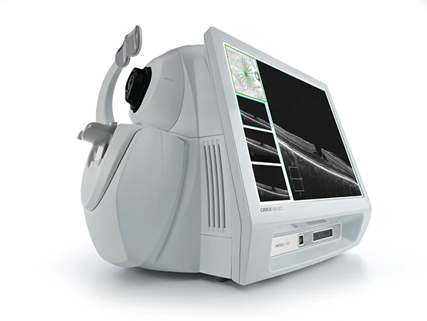
Zeiss hd oct for advanced glaucoma imaging
1. OCT ANFL
2. OCT GCC
3. OCT PACHYMETRY
4. OCT GONIOSCOPY

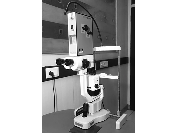
Yag laser for peripheral iridotomy.

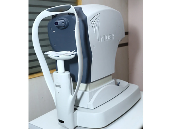
Non contact tonometry

Zeiss fundus camera for optic nerve imaging

Zeiss humphrey’s visual field analyser is the gold standard device for early detection and monitoring progression of glaucoma
Frequently asked questions on glaucoma treatment or kala motiya
Unfortunately, most cases of glaucoma do not occur with readily noticeable symptoms. However, the presence of the following warning signs, indicates that you need a thorough examination by an eye doctor:
• Unusual trouble adjusting to dark rooms
• Difficulty in focussing near/distant objects
• Squinting or blinking due to unusual sensitivity to light/glare
• Red-rimmed, encrusted or swollen lids
• Recurrent pain in or around eyes
• Dark spot at the centre of viewing
• Lines and edges appear distorted or wavy
• Excess tearing or "watery eyes"
• Double vision
The following may be indications of potentially serious problems that might require emergency medical attention:
• Sudden loss ofvision in one eye
• Sudden hazy or blurred vision
• Flashes oflight or black spots
• Halos or rainbows around light
The symptoms listed above may not necessarily mean that you have glaucoma. However, if you experience one or more of these symptoms, contact your eye doctor for a complete exam.
The most important risk factors include:
• Age (above 45 yrs.)
• Elevated eye pressure
• Thin cornea
• Family history of glaucoma
• Nearsightedness
• Past injury to the eyes
• Steroid use
• A history of severe anemia or shock
• Conditions affecting flow of blood like diabetes, migraine, blood pressure
Your ophthalmologist will weigh all these factors before deciding for treatment for glaucoma or a regular monitoring as a glaucoma suspect to detect the early signs of damage to the optic nerve.
Open-angle glaucoma
It is the most common form of glaucoma. The *open* drainage angle of the eye may get blocked leading to gradual increased eye pressure. If this increased pressure results in optic nerve damage, it is known as chronic open-angle glaucoma. The optic nerve damage & vision loss usually occurs so gradually and painlessly that you are not aware of trouble until the optic nerve is already badly damaged
Closed angle glaucoma / Angle-closure glaucoma
It results when the drainage angle of the eye narrows and becomes completely blocked. In the eye, the iris may close off the drainage angle and cause a dangerously high eye pressure. This complete block results in building up of pressure and is called as acute angle-closure glaucoma. The symptoms include severe eye pain, blurred vision, headache, rainbow haloes around lights, nausea and vomiting. When the drainage angle of the eye gradually becomes completely blocked, pressure builds up gradually, results in chronic angle-closure glaucoma.
Other types of glaucoma
Not all types of glaucoma are characterized by eye pressures. In normal-tension or low-tension glaucoma, the optic nerve suffers damage with the resulting visual field loss even though normal eye pressures are maintained.
1. Exfoliation syndrome
A common form of open-angle glaucoma that results where there is a build-up of abnormal, whitish material on the lens and drainage angle of the eye. This causes increased eye pressure.
2. Due to injuries or disease ofeyes
Other types of glaucoma may be caused by injuries to the eye, tumors, and other eye diseases.
3. Congenital Glaucoma
A rare type of glaucoma can even be present in children at birth.
Regular eye examinations by your ophthalmologist are the best way to detect glaucoma. Your ophthalmologist will
• Measure your eye pressure (tonometry);
• Inspect the drainage angle of your eye (gonioscopy);
• Evaluate your optic nerve (ophthalmolscopy);
• Test the visual field of each eye (perimetry).
Optic nerve evaluation and visual field testing are performed at regular intervals to monitor the effects of glaucoma. The objective is to determine if glaucoma damage has progressed over time.
The treatment for glaucoma depends upon the nature and severity of each case. Eye drops, pills, laser procedures, and surgical operations are used to prevent or slow further damage from occurring. Regular eye examinations are very important to detect progression and to prevent vision loss. Because glaucoma can worsen without your being aware of it, your treatment will likely need to be changed over time to achieve a lower "target eye pressure."
Medicines
Glaucoma is often treated with eye drops taken regularly several times a day, sometimes in combination with pills. These medications will alter the circulation of eye fluid and lower eye pressure.
Laser Surgery
Laser surgery is also effective for glaucoma treatment.
Trabeculoplasty is laser treatment to enhance the eye drainage function to control eye pressure within the eye when treating open-angle glaucoma. Iridotomy is laser treatment to create tiny holes in the iris to improve the flow of eye fluid to the drain when treating narrow angle glaucoma.
Operative Surgery
In some patients, surgical intervention may be required. Your Ophthalmologist is the best person to guide you in this regard.


