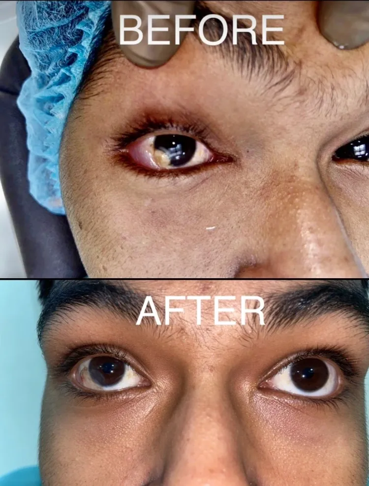
Introduction:
The human eye is a complex organ, and any abnormalities that affect its structure can have a significant impact on vision and overall eye health. One such condition is Limbal Corneal Dermoid, a rare but noteworthy eye disorder that requires attention and proper management. In this blog post, we will explore the causes, symptoms, and treatment options for Limbal Corneal Dermoid.
What is Limbal Corneal Dermoid?
Limbal Corneal Dermoid is a congenital condition characterized by the presence of noncancerous skin-like tissue in the cornea, specifically at the limbal region. The limbus is the border between the cornea (the clear front part of the eye) and the sclera (the white outer layer of the eye). Dermoids typically consist of various tissues, such as skin, hair follicles, and sweat glands, and can vary in size and appearance.
Causes:
The exact cause of Limbal Corneal Dermoid is unknown. It is believed to be a result of abnormal embryonic development during pregnancy. However, no specific genetic or environmental factors have been identified as direct causes of this condition.
Symptoms:
The symptoms of Limbal Corneal Dermoid can vary depending on the size and location of the dermoid. Common signs and symptoms include:
1. White or yellowish elevated mass on the cornea or limbal area.
2. Visible hair or hair-like structures on the cornea.
3. Irritation or redness in the affected eye.
4. Sensation of a foreign body or discomfort.
5. Blurred or distorted vision if the dermoid affects the central visual axis.
It’s important to note that Limbal Corneal Dermoid may be present at birth or appear later in childhood.
Diagnosis and Treatment:
If Limbal Corneal Dermoid is suspected, an ophthalmologist will perform a comprehensive eye examination, which may include the use of specialized imaging techniques such as corneal topography or ultrasound. The diagnosis is typically confirmed by observing the presence of abnormal tissue on the cornea.
Treatment options for Limbal Corneal Dermoid depend on several factors, including the size and location of the dermoid, as well as the impact on vision and overall eye health. In some cases, if the dermoid is small and not causing any significant visual disturbances or discomfort, close observation and regular eye examinations may be recommended.
However, if the dermoid is large, affects vision, or causes significant discomfort, surgical intervention may be necessary. Surgical options include excision of the dermoid with tissue grafting or the use of advanced techniques such as laser ablation or cryotherapy to reduce the size of the dermoid. The choice of treatment depends on individual circumstances and should be discussed with an experienced eye surgeon.
Conclusion:
Limbal Corneal Dermoid is a rare congenital eye condition characterized by the presence of skin-like tissue on the cornea’s limbal region. While the exact cause is unknown, proper diagnosis and management are crucial to ensure optimal eye health and visual function. If you suspect you or your child may have Limbal Corneal Dermoid, consult an ophthalmologist for a comprehensive evaluation and personalized treatment recommendations. Early detection and appropriate intervention can significantly improve outcomes and enhance quality of life for those affected by this condition.


